Achieve Mastery of Medical Concepts

Study for medical school and boards with Lecturio

Blood Group Systems

There are 29 blood group systems, among which the ABO group is the most important. Blood groups are determined by antigens that are surface markers on red blood cells Red blood cells Erythrocytes, or red blood cells (RBCs), are the most abundant cells in the blood. While erythrocytes in the fetus are initially produced in the yolk sac then the liver, the bone marrow eventually becomes the main site of production. Erythrocytes: Histology ( RBCs RBCs Erythrocytes, or red blood cells (RBCs), are the most abundant cells in the blood. While erythrocytes in the fetus are initially produced in the yolk sac then the liver, the bone marrow eventually becomes the main site of production. Erythrocytes: Histology) and composed of proteins Proteins Linear polypeptides that are synthesized on ribosomes and may be further modified, crosslinked, cleaved, or assembled into complex proteins with several subunits. The specific sequence of amino acids determines the shape the polypeptide will take, during protein folding, and the function of the protein. Energy Homeostasis and carbohydrates Carbohydrates A class of organic compounds composed of carbon, hydrogen, and oxygen in a ratio of cn(H2O)n. The largest class of organic compounds, including starch; glycogen; cellulose; polysaccharides; and simple monosaccharides. Basics of Carbohydrates. Antigens are also found on platelets Platelets Platelets are small cell fragments involved in hemostasis. Thrombopoiesis takes place primarily in the bone marrow through a series of cell differentiation and is influenced by several cytokines. Platelets are formed after fragmentation of the megakaryocyte cytoplasm. Platelets: Histology, leukocytes Leukocytes White blood cells. These include granular leukocytes (basophils; eosinophils; and neutrophils) as well as non-granular leukocytes (lymphocytes and monocytes). White Myeloid Cells: Histology, and tissue cells as well as in soluble form in body secretions such as breast milk, seminal fluid, saliva Saliva The clear, viscous fluid secreted by the salivary glands and mucous glands of the mouth. It contains mucins, water, organic salts, and ptyalin. Salivary Glands: Anatomy, sweat, gastric secretions Gastric secretions Gastrointestinal Secretions, urine, and amniotic fluid Amniotic fluid A clear, yellowish liquid that envelopes the fetus inside the sac of amnion. In the first trimester, it is likely a transudate of maternal or fetal plasma. In the second trimester, amniotic fluid derives primarily from fetal lung and kidney. Cells or substances in this fluid can be removed for prenatal diagnostic tests (amniocentesis). Placenta, Umbilical Cord, and Amniotic Cavity. Individuals will naturally develop antibodies Antibodies Immunoglobulins (Igs), also known as antibodies, are glycoprotein molecules produced by plasma cells that act in immune responses by recognizing and binding particular antigens. The various Ig classes are IgG (the most abundant), IgM, IgE, IgD, and IgA, which differ in their biologic features, structure, target specificity, and distribution. Immunoglobulins: Types and Functions (Abs) against the ABO antigens they do not have. For this reason, determining an individual’s blood group is important prior to any blood product transfusion and prior to donating or receiving an organ transplant. If appropriate matching does not happen, a massive activation of the immune system Immune system The body's defense mechanism against foreign organisms or substances and deviant native cells. It includes the humoral immune response and the cell-mediated response and consists of a complex of interrelated cellular, molecular, and genetic components. Primary Lymphatic Organs and coagulation cascade Coagulation cascade The coagulation cascade is a series of reactions that ultimately generates a strong, cross-linked fibrin clot. Hemostasis will ensue, leading to shock Shock Shock is a life-threatening condition associated with impaired circulation that results in tissue hypoxia. The different types of shock are based on the underlying cause: distributive (↑ cardiac output (CO), ↓ systemic vascular resistance (SVR)), cardiogenic (↓ CO, ↑ SVR), hypovolemic (↓ CO, ↑ SVR), obstructive (↓ CO), and mixed. Types of Shock, organ failure, and even death.

Last updated: Dec 19, 2022

Editorial responsibility: Stanley Oiseth, Lindsay Jones, Evelin Maza

ABO Blood Group

Antigens

There are 4 common blood groups in the ABO system: A, B, AB, and O.
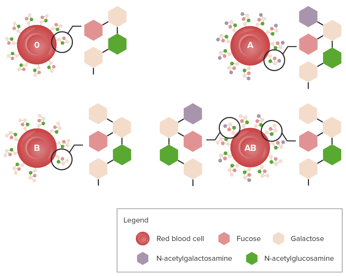
  • These blood groups are defined by the presence of specific antigens on RBCs RBCs Erythrocytes, or red blood cells (RBCs), are the most abundant cells in the blood. While erythrocytes in the fetus are initially produced in the yolk sac then the liver, the bone marrow eventually becomes the main site of production. Erythrocytes: Histology.
    • Blood group A: antigen Antigen Substances that are recognized by the immune system and induce an immune reaction. Vaccination A
    • Blood group B: antigen Antigen Substances that are recognized by the immune system and induce an immune reaction. Vaccination B
    • Blood group AB: antigens A and B
    • Blood group O: no antigens
  • A and B antigens consist of specific carbohydrate sugars:
    • N-acetylgalactosamine for the A antigen Antigen Substances that are recognized by the immune system and induce an immune reaction. Vaccination 
    • D-galactose for the B antigen Antigen Substances that are recognized by the immune system and induce an immune reaction. Vaccination 
    • Both of these sugars bind BIND Hyperbilirubinemia of the Newborn to and modify the H antigen H antigen Salmonella on the surface of RBCs RBCs Erythrocytes, or red blood cells (RBCs), are the most abundant cells in the blood. While erythrocytes in the fetus are initially produced in the yolk sac then the liver, the bone marrow eventually becomes the main site of production. Erythrocytes: Histology.
    • If the H antigen H antigen Salmonella is left unmodified, the resulting blood group is O. 
  • There are over 80 ABO alleles: The most common are A1, A2, B1, O1, O1v, and O2.
Diagram showing the carbohydrate chains that determine the abo blood group

Diagram showing the carbohydrate chains that determine the ABO blood group

Image by Lecturio.

Inheritance

Blood-type frequencies vary in different racial/ethnic groups. The ABO blood type is inherited in an autosomal codominant fashion:

  • The A and B alleles are codominant.
  • The O allele Allele Variant forms of the same gene, occupying the same locus on homologous chromosomes, and governing the variants in production of the same gene product. Basic Terms of Genetics is recessive.
Table: Inheritance pattern for ABO blood group
Mother A Mother B Mother O
Father A AA AA Amyloidosis AB AO
Father B BA BB BO
Father O OA OA Osteoarthritis (OA) is the most common form of arthritis, and is due to cartilage destruction and changes of the subchondral bone. The risk of developing this disorder increases with age, obesity, and repetitive joint use or trauma. Patients develop gradual joint pain, stiffness lasting < 30 minutes, and decreased range of motion. Osteoarthritis OB OO
Table: Alleles and blood types
Alleles Blood type
A + A A
A + O A
A + B AB
B + B B
B + O B
O + O O
The dominant alleles are A and B; when both of them are present, the individual has the blood type AB. The recessive allele is O.

Antibodies Antibodies Immunoglobulins (Igs), also known as antibodies, are glycoprotein molecules produced by plasma cells that act in immune responses by recognizing and binding particular antigens. The various Ig classes are IgG (the most abundant), IgM, IgE, IgD, and IgA, which differ in their biologic features, structure, target specificity, and distribution. Immunoglobulins: Types and Functions

Individuals will naturally develop antibodies Antibodies Immunoglobulins (Igs), also known as antibodies, are glycoprotein molecules produced by plasma cells that act in immune responses by recognizing and binding particular antigens. The various Ig classes are IgG (the most abundant), IgM, IgE, IgD, and IgA, which differ in their biologic features, structure, target specificity, and distribution. Immunoglobulins: Types and Functions (Abs) against the ABO antigens they do not have:

  • Individuals with blood group A have anti-B Ab.
  • Individuals with blood group B have anti-A Ab.
  • Individuals with blood group O will have both anti-A and anti-B Abs.

These Abs can elicit a hemolytic response upon encountering their respective antigen Antigen Substances that are recognized by the immune system and induce an immune reaction. Vaccination:

  • An ABO-incompatible blood transfusion can be fatal.
  • A universal RBC donor has type O blood.
  • A universal plasma Plasma The residual portion of blood that is left after removal of blood cells by centrifugation without prior blood coagulation. Transfusion Products donor has type AB blood.

Associated risks

Blood groups are associated with differential risk to certain diseases. Blood group O may have ↓ risk for: 
  • Pancreatic cancer 
  • Thromboembolic disease
  • Malaria Malaria Malaria is an infectious parasitic disease affecting humans and other animals. Most commonly transmitted via the bite of a female Anopheles mosquito infected with microorganisms of the Plasmodium genus. Patients present with fever, chills, myalgia, headache, and diaphoresis. Plasmodium/Malaria
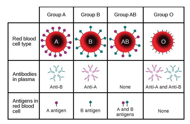
Blood-type antigens

Diagram showing that a person may express A, B, AB, or no antigen at all:
The diagram also describes the type of antibody produced by the carrier.

Image: “ABO blood group antigens present on red blood cells and IgM antibodies present in the serum” by InvictaHOG. License: Public Domain

Rh Factor

Definition

Rh factor (Rhesus factor) is an RBC surface antigen Antigen Substances that are recognized by the immune system and induce an immune reaction. Vaccination.

  • Encoded by the 2 genes Genes A category of nucleic acid sequences that function as units of heredity and which code for the basic instructions for the development, reproduction, and maintenance of organisms. DNA Types and Structure RHD and RHCE:
    • Polymorphism Polymorphism The regular and simultaneous occurrence in a single interbreeding population of two or more discontinuous genotypes. The concept includes differences in genotypes ranging in size from a single nucleotide site to large nucleotide sequences visible at a chromosomal level. Basic Terms of Genetics: Numerous genetic rearrangements produce distinct Rh antigens.
    • 50 Rh group antigens (5 are important)
  • 85% of people carry the D allele Allele Variant forms of the same gene, occupying the same locus on homologous chromosomes, and governing the variants in production of the same gene product. Basic Terms of Genetics: They are Rh+. 
  • 15% do not carry it: They are Rh–.
  • Rh– persons do not carry anti-Rh Ab unless they are exposed to Rh+ RBCs RBCs Erythrocytes, or red blood cells (RBCs), are the most abundant cells in the blood. While erythrocytes in the fetus are initially produced in the yolk sac then the liver, the bone marrow eventually becomes the main site of production. Erythrocytes: Histology.

Rh incompatibility Rh Incompatibility Hemolytic Disease of the Fetus and Newborn

Anti-Rh Abs occur when:

  • Rh+ blood is transfused into Rh– person:
    • Causes an acute hemolytic reaction
    • Results in a mild hemolytic anemia Hemolytic Anemia Hemolytic anemia (HA) is the term given to a large group of anemias that are caused by the premature destruction/hemolysis of circulating red blood cells (RBCs). Hemolysis can occur within (intravascular hemolysis) or outside the blood vessels (extravascular hemolysis). Hemolytic Anemia
  • Rh– pregnant mother is exposed to Rh+ fetal RBCs RBCs Erythrocytes, or red blood cells (RBCs), are the most abundant cells in the blood. While erythrocytes in the fetus are initially produced in the yolk sac then the liver, the bone marrow eventually becomes the main site of production. Erythrocytes: Histology:
    • Secondary to fetomaternal hemorrhage from abortion Abortion Expulsion of the product of fertilization before completing the term of gestation and without deliberate interference. Spontaneous Abortion, trauma, invasive obstetric procedures, or normal delivery
    • Mother produces Abs against the baby’s RBCs RBCs Erythrocytes, or red blood cells (RBCs), are the most abundant cells in the blood. While erythrocytes in the fetus are initially produced in the yolk sac then the liver, the bone marrow eventually becomes the main site of production. Erythrocytes: Histology
    • Abs are IgG IgG The major immunoglobulin isotype class in normal human serum. There are several isotype subclasses of igg, for example, igg1, igg2a, and igg2b. Hypersensitivity Pneumonitis and cross the placenta Placenta A highly vascularized mammalian fetal-maternal organ and major site of transport of oxygen, nutrients, and fetal waste products. It includes a fetal portion (chorionic villi) derived from trophoblasts and a maternal portion (decidua) derived from the uterine endometrium. The placenta produces an array of steroid, protein and peptide hormones (placental hormones). Placenta, Umbilical Cord, and Amniotic Cavity
    • Abs can cause hemolysis to the fetus (also called hemolytic disease of the newborn Newborn An infant during the first 28 days after birth. Physical Examination of the Newborn or erythroblastosis fetalis Erythroblastosis fetalis Hemolytic disease of the fetus and newborn (HDFN), also known as erythroblastosis fetalis, is caused by maternal IgG antibody destruction of the fetal RBCs. Rhesus (Rh) blood group incompatibility (frequently triggered by D antigen) and ABO incompatibility are common causes. Hemolytic Disease of the Fetus and Newborn).
    • Anti-D Ig Ig X-linked Agammaglobulinemia is administered to sensitized women after pregnancy Pregnancy The status during which female mammals carry their developing young (embryos or fetuses) in utero before birth, beginning from fertilization to birth. Pregnancy: Diagnosis, Physiology, and Care to avoid this reaction.

If fetus is Rh+ and the mother Rh–, serial monitoring during pregnancy Pregnancy The status during which female mammals carry their developing young (embryos or fetuses) in utero before birth, beginning from fertilization to birth. Pregnancy: Diagnosis, Physiology, and Care is needed and is accomplished by:
  • Serial indirect Coombs test in mother
  • Measurement of fetal blood flow velocity Flow velocity Vascular Resistance, Flow, and Mean Arterial Pressure in middle cerebral artery Middle cerebral artery The largest of the cerebral arteries. It trifurcates into temporal, frontal, and parietal branches supplying blood to most of the parenchyma of these lobes in the cerebral cortex. These are the areas involved in motor, sensory, and speech activities. Cerebrovascular System: Anatomy by Doppler Doppler Ultrasonography applying the doppler effect, with frequency-shifted ultrasound reflections produced by moving targets (usually red blood cells) in the bloodstream along the ultrasound axis in direct proportion to the velocity of movement of the targets, to determine both direction and velocity of blood flow. Ultrasound (Sonography)
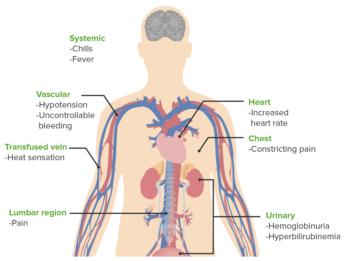
Signs and symptoms of acute hemolytic transfusion reactions

Signs and symptoms of acute hemolytic transfusion reactions

Image by Lecturio.

Other Blood Groups

Kell system

  • 3rd most potent immunogenic antigen Antigen Substances that are recognized by the immune system and induce an immune reaction. Vaccination after ABO and Rh system
  • 25 Kell antigens have been discovered.
  • Defined by the immune Ab anti-K
  • Anti-K Ab causes:
    • Severe hemolytic disease of the fetus Hemolytic disease of the fetus Hemolytic disease of the fetus and newborn (HDFN), also known as erythroblastosis fetalis, is caused by maternal IgG antibody destruction of the fetal RBCs. Rhesus (Rh) blood group incompatibility (frequently triggered by D antigen) and ABO incompatibility are common causes. Hemolytic Disease of the Fetus and Newborn and newborn Newborn An infant during the first 28 days after birth. Physical Examination of the Newborn
    • Hemolytic transfusion reactions Transfusion reactions Transfusion-related complications occur during or after a blood product is given. These complications can be classified as immunologic, non-immunologic and acute, and delayed. Non-immunologic reactions are caused by the transmission of disease in blood products, and immunologic reactions are antigen-antibody-mediated. Transfusion Reactions

Duffy system

  • Duffy antigen Antigen Substances that are recognized by the immune system and induce an immune reaction. Vaccination was named after a patient with hemophilia Hemophilia The hemophilias are a group of inherited, or sometimes acquired, disorders of secondary hemostasis due to deficiency of specific clotting factors. Hemophilia A is a deficiency of factor VIII, hemophilia B a deficiency of factor IX, and hemophilia C a deficiency of factor XI. Patients present with bleeding events that may be spontaneous or associated with minor or major trauma. Hemophilia who received many transfusions and developed antibodies Antibodies Immunoglobulins (Igs), also known as antibodies, are glycoprotein molecules produced by plasma cells that act in immune responses by recognizing and binding particular antigens. The various Ig classes are IgG (the most abundant), IgM, IgE, IgD, and IgA, which differ in their biologic features, structure, target specificity, and distribution. Immunoglobulins: Types and Functions to the antigen Antigen Substances that are recognized by the immune system and induce an immune reaction. Vaccination.
  • Also known as Fy glycoprotein
  • 2 alleles exist:
    • Fya
    • Fyb
  • There are 4 possible phenotypes:
    • Fy(a+b–)
    • Fy(a+b+)
    • Fy(a–b+)
    • Fy(a–b–)
  • Nonspecific receptor Receptor Receptors are proteins located either on the surface of or within a cell that can bind to signaling molecules known as ligands (e.g., hormones) and cause some type of response within the cell. Receptors for chemokines Chemokines Class of pro-inflammatory cytokines that have the ability to attract and activate leukocytes. They can be divided into at least three structural branches: c; cc; and cxc; according to variations in a shared cysteine motif. Adaptive Cell-mediated Immunity and the malaria Malaria Malaria is an infectious parasitic disease affecting humans and other animals. Most commonly transmitted via the bite of a female Anopheles mosquito infected with microorganisms of the Plasmodium genus. Patients present with fever, chills, myalgia, headache, and diaphoresis. Plasmodium/Malaria parasite Plasmodium Plasmodium A genus of protozoa that comprise the malaria parasites of mammals. Four species infect humans (although occasional infections with primate malarias may occur). These are plasmodium falciparum; plasmodium malariae; plasmodium ovale, and plasmodium vivax. Species causing infection in vertebrates other than man include: plasmodium berghei; plasmodium chabaudi; p. Vinckei, and plasmodium yoelii in rodents; p. Brasilianum, plasmodium cynomolgi; and plasmodium knowlesi in monkeys; and plasmodium gallinaceum in chickens. Antimalarial Drugs vivax
  • Associated Abs are IgG IgG The major immunoglobulin isotype class in normal human serum. There are several isotype subclasses of igg, for example, igg1, igg2a, and igg2b. Hypersensitivity Pneumonitis and can cause hemolytic transfusion reactions Transfusion reactions Transfusion-related complications occur during or after a blood product is given. These complications can be classified as immunologic, non-immunologic and acute, and delayed. Non-immunologic reactions are caused by the transmission of disease in blood products, and immunologic reactions are antigen-antibody-mediated. Transfusion Reactions.

Kidd system

  • Kidd antigen Antigen Substances that are recognized by the immune system and induce an immune reaction. Vaccination (Jk antigen Antigen Substances that are recognized by the immune system and induce an immune reaction. Vaccination) acts as an urea Urea A compound formed in the liver from ammonia produced by the deamination of amino acids. It is the principal end product of protein catabolism and constitutes about one half of the total urinary solids. Urea Cycle transporter in RBC and renal endothelium Endothelium A layer of epithelium that lines the heart, blood vessels (vascular endothelium), lymph vessels (lymphatic endothelium), and the serous cavities of the body. Arteries: Histology.
  • 3 antigens have been identified to date:
    • Jka
    • Jkb
    • JK3
  • Kidd Ab can cause severe transfusion reactions Transfusion reactions Transfusion-related complications occur during or after a blood product is given. These complications can be classified as immunologic, non-immunologic and acute, and delayed. Non-immunologic reactions are caused by the transmission of disease in blood products, and immunologic reactions are antigen-antibody-mediated. Transfusion Reactions.

Complications of Blood Transfusion

Complications of blood transfusion can be acute (minutes to hours) or delayed (days to months).
Acute

  • Hemolytic reaction:
    • Immediate or within 24 hours of transfusion:
      • Due to IgM IgM A class of immunoglobulin bearing mu chains (immunoglobulin mu-chains). Igm can fix complement. The name comes from its high molecular weight and originally being called a macroglobulin. Immunoglobulins: Types and Functions antibodies Antibodies Immunoglobulins (Igs), also known as antibodies, are glycoprotein molecules produced by plasma cells that act in immune responses by recognizing and binding particular antigens. The various Ig classes are IgG (the most abundant), IgM, IgE, IgD, and IgA, which differ in their biologic features, structure, target specificity, and distribution. Immunoglobulins: Types and Functions against RBCs RBCs Erythrocytes, or red blood cells (RBCs), are the most abundant cells in the blood. While erythrocytes in the fetus are initially produced in the yolk sac then the liver, the bone marrow eventually becomes the main site of production. Erythrocytes: Histology
      • Typically of the ABO group
    • Delayed: IgG IgG The major immunoglobulin isotype class in normal human serum. There are several isotype subclasses of igg, for example, igg1, igg2a, and igg2b. Hypersensitivity Pneumonitis antibodies Antibodies Immunoglobulins (Igs), also known as antibodies, are glycoprotein molecules produced by plasma cells that act in immune responses by recognizing and binding particular antigens. The various Ig classes are IgG (the most abundant), IgM, IgE, IgD, and IgA, which differ in their biologic features, structure, target specificity, and distribution. Immunoglobulins: Types and Functions are not present during transfusion but develop later. 
    • Nonimmune:
      • Due to aged RBCs RBCs Erythrocytes, or red blood cells (RBCs), are the most abundant cells in the blood. While erythrocytes in the fetus are initially produced in the yolk sac then the liver, the bone marrow eventually becomes the main site of production. Erythrocytes: Histology
      • Improper storage
      • Transfusion along noncompatible medications 
      • Transfusion through small-bore IV tubes
  • Infection:
    • Bacterial
    • Viral: HIV HIV Anti-HIV Drugs, hepatitis C Hepatitis C Hepatitis C is an infection of the liver caused by the hepatitis C virus (HCV). The infection can be transmitted through infectious blood or body fluids and may be transmitted during childbirth or through IV drug use or sexual intercourse. Hepatitis C virus can cause both acute and chronic hepatitis, ranging from a mild to a serious, lifelong illness including liver cirrhosis and hepatocellular carcinoma (HCC). Hepatitis C Virus virus Virus Viruses are infectious, obligate intracellular parasites composed of a nucleic acid core surrounded by a protein capsid. Viruses can be either naked (non-enveloped) or enveloped. The classification of viruses is complex and based on many factors, including type and structure of the nucleoid and capsid, the presence of an envelope, the replication cycle, and the host range. Virology, hepatitis B Hepatitis B Hepatitis B virus (HBV) is a partially double-stranded DNA virus, which belongs to the Orthohepadnavirus genus and the Hepadnaviridae family. Most individuals with acute HBV infection are asymptomatic or have mild, self-limiting symptoms. Chronic infection can be asymptomatic or create hepatic inflammation, leading to liver cirrhosis and hepatocellular carcinoma (HCC). Hepatitis B Virus virus Virus Viruses are infectious, obligate intracellular parasites composed of a nucleic acid core surrounded by a protein capsid. Viruses can be either naked (non-enveloped) or enveloped. The classification of viruses is complex and based on many factors, including type and structure of the nucleoid and capsid, the presence of an envelope, the replication cycle, and the host range. Virology, human T-lymphotropic virus Virus Viruses are infectious, obligate intracellular parasites composed of a nucleic acid core surrounded by a protein capsid. Viruses can be either naked (non-enveloped) or enveloped. The classification of viruses is complex and based on many factors, including type and structure of the nucleoid and capsid, the presence of an envelope, the replication cycle, and the host range. Virology 1 or 2
    • Very rare due to screening Screening Preoperative Care/treatment protocols for donated blood products
  • Allergic reaction:
    • Mild: hives Hives Urticaria is raised, well-circumscribed areas (wheals) of edema (swelling) and erythema (redness) involving the dermis and epidermis with associated pruritus (itch). Urticaria is not a single disease but rather is a reaction pattern representing cutaneous mast cell degranulation. Urticaria (Hives), pruritus Pruritus An intense itching sensation that produces the urge to rub or scratch the skin to obtain relief. Atopic Dermatitis (Eczema)
    • Severe, leading to anaphylaxis Anaphylaxis An acute hypersensitivity reaction due to exposure to a previously encountered antigen. The reaction may include rapidly progressing urticaria, respiratory distress, vascular collapse, systemic shock, and death. Type I Hypersensitivity Reaction:
      • Hypotension Hypotension Hypotension is defined as low blood pressure, specifically < 90/60 mm Hg, and is most commonly a physiologic response. Hypotension may be mild, serious, or life threatening, depending on the cause. Hypotension
      • Bronchospasm Bronchospasm Asthma Drugs 
      • GI symptoms ( nausea Nausea An unpleasant sensation in the stomach usually accompanied by the urge to vomit. Common causes are early pregnancy, sea and motion sickness, emotional stress, intense pain, food poisoning, and various enteroviruses. Antiemetics, abdominal pain Abdominal Pain Acute Abdomen)
      • Systemic organ failure
    • Due to sensitization to antigens in the donor unit, especially plasma Plasma The residual portion of blood that is left after removal of blood cells by centrifugation without prior blood coagulation. Transfusion Products proteins Proteins Linear polypeptides that are synthesized on ribosomes and may be further modified, crosslinked, cleaved, or assembled into complex proteins with several subunits. The specific sequence of amino acids determines the shape the polypeptide will take, during protein folding, and the function of the protein. Energy Homeostasis
  • Transfusion-related acute lung injury Transfusion-related acute lung injury A rare but serious transfusion-related reaction in which fluid builds up in the lungs unrelated to excessively high infusion rate and/or volume (transfusion-associated circulatory overload). Signs of transfusion-related acute lung injury include pulmonary secretions; hypotension; fever; dyspnea; tachypnea; tachycardia; and cyanosis. Transfusion Reactions (TRALI):
  • Acute febrile nonhemolytic reaction: 
    • Body temperature Body Temperature The measure of the level of heat of a human or animal. Heatstroke by 1°F (-17°C) within 24 hours after blood transfusion
    • More frequent with:
    • Due to release of Ab-mediated endogenous pyrogens Pyrogens Substances capable of increasing body temperature and cause fever and may be used for fever therapy. They may be of microbial origin, often polysaccharides, and may contaminate distilled water. Fever and cytokines Cytokines Non-antibody proteins secreted by inflammatory leukocytes and some non-leukocytic cells, that act as intercellular mediators. They differ from classical hormones in that they are produced by a number of tissue or cell types rather than by specialized glands. They generally act locally in a paracrine or autocrine rather than endocrine manner. Adaptive Immune Response
  • Circulatory overload: 
    • Due to rapid transfusion of large volume of blood products
    • ↑ Risk:
      • Underlying cardiopulmonary disease
      • Renal failure Renal failure Conditions in which the kidneys perform below the normal level in the ability to remove wastes, concentrate urine, and maintain electrolyte balance; blood pressure; and calcium metabolism. Renal insufficiency can be classified by the degree of kidney damage (as measured by the level of proteinuria) and reduction in glomerular filtration rate. Crush Syndrome
      • Chronic anemia Anemia Anemia is a condition in which individuals have low Hb levels, which can arise from various causes. Anemia is accompanied by a reduced number of RBCs and may manifest with fatigue, shortness of breath, pallor, and weakness. Subtypes are classified by the size of RBCs, chronicity, and etiology. Anemia: Overview and Types
      • Elderly patients Patients Individuals participating in the health care system for the purpose of receiving therapeutic, diagnostic, or preventive procedures. Clinician–Patient Relationship 
    • Signs and symptoms include:
      • ↑ HR, dyspnea Dyspnea Dyspnea is the subjective sensation of breathing discomfort. Dyspnea is a normal manifestation of heavy physical or psychological exertion, but also may be caused by underlying conditions (both pulmonary and extrapulmonary). Dyspnea, cough 
      • Central venous pressure Central venous pressure The blood pressure in the central large veins of the body. It is distinguished from peripheral venous pressure which occurs in an extremity. Central Venous Catheter, widened pulse pressure 
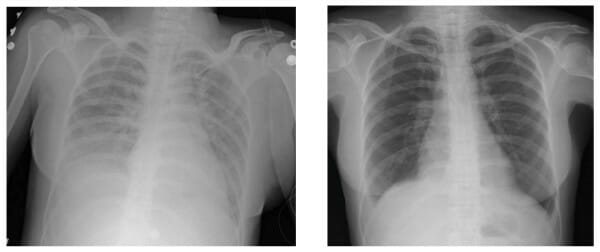
      • Chest radiography shows cardiomegaly Cardiomegaly Enlargement of the heart, usually indicated by a cardiothoracic ratio above 0. 50. Heart enlargement may involve the right, the left, or both heart ventricles or heart atria. Cardiomegaly is a nonspecific symptom seen in patients with chronic systolic heart failure (heart failure) or several forms of cardiomyopathies. Ebstein’s Anomaly and pulmonary edema Pulmonary edema Pulmonary edema is a condition caused by excess fluid within the lung parenchyma and alveoli as a consequence of a disease process. Based on etiology, pulmonary edema is classified as cardiogenic or noncardiogenic. Patients may present with progressive dyspnea, orthopnea, cough, or respiratory failure. Pulmonary Edema.

Delayed

  • Iron overload Iron overload An excessive accumulation of iron in the body due to a greater than normal absorption of iron from the gastrointestinal tract or from parenteral injection. This may arise from idiopathic hemochromatosis, excessive iron intake, chronic alcoholism, certain types of refractory anemia, or transfusional hemosiderosis. Hereditary Hemochromatosis:
    • In patients Patients Individuals participating in the health care system for the purpose of receiving therapeutic, diagnostic, or preventive procedures. Clinician–Patient Relationship who receive blood long term and regularly
    • 1 unit of RBC = 200 mg of iron Iron A metallic element with atomic symbol fe, atomic number 26, and atomic weight 55. 85. It is an essential constituent of hemoglobins; cytochromes; and iron-binding proteins. It plays a role in cellular redox reactions and in the transport of oxygen. Trace Elements
    • Iron Iron A metallic element with atomic symbol fe, atomic number 26, and atomic weight 55. 85. It is an essential constituent of hemoglobins; cytochromes; and iron-binding proteins. It plays a role in cellular redox reactions and in the transport of oxygen. Trace Elements accumulation can occur and is toxic for tissues and organs.
    • Management: iron Iron A metallic element with atomic symbol fe, atomic number 26, and atomic weight 55. 85. It is an essential constituent of hemoglobins; cytochromes; and iron-binding proteins. It plays a role in cellular redox reactions and in the transport of oxygen. Trace Elements chelation
  • Graft-versus-host disease Graft-versus-host disease The clinical entity characterized by anorexia, diarrhea, loss of hair, leukopenia, thrombocytopenia, growth retardation, and eventual death brought about by the graft vs host reaction. Transfusion Reactions:
    • Due to proliferation of donor’s lymphocytes Lymphocytes Lymphocytes are heterogeneous WBCs involved in immune response. Lymphocytes develop from the bone marrow, starting from hematopoietic stem cells (HSCs) and progressing to common lymphoid progenitors (CLPs). B and T lymphocytes and natural killer (NK) cells arise from the lineage. Lymphocytes: Histology, which attack recipient’s tissues and organs
    • Symptoms include:
      • Rash Rash Rocky Mountain Spotted Fever, fever Fever Fever is defined as a measured body temperature of at least 38°C (100.4°F). Fever is caused by circulating endogenous and/or exogenous pyrogens that increase levels of prostaglandin E2 in the hypothalamus. Fever is commonly associated with chills, rigors, sweating, and flushing of the skin. Fever, diarrhea Diarrhea Diarrhea is defined as ≥ 3 watery or loose stools in a 24-hour period. There are a multitude of etiologies, which can be classified based on the underlying mechanism of disease. The duration of symptoms (acute or chronic) and characteristics of the stools (e.g., watery, bloody, steatorrheic, mucoid) can help guide further diagnostic evaluation. Diarrhea, liver Liver The liver is the largest gland in the human body. The liver is found in the superior right quadrant of the abdomen and weighs approximately 1.5 kilograms. Its main functions are detoxification, metabolism, nutrient storage (e.g., iron and vitamins), synthesis of coagulation factors, formation of bile, filtration, and storage of blood. Liver: Anatomy dysfunction, and pancytopenia Pancytopenia Deficiency of all three cell elements of the blood, erythrocytes, leukocytes and platelets. Aplastic Anemia 
      • Symptoms occur 1–6 weeks after transfusion
    • Fatal in > 90% of cases
    • ↑ Risk in:
      • Immunocompromised immunocompromised A human or animal whose immunologic mechanism is deficient because of an immunodeficiency disorder or other disease or as the result of the administration of immunosuppressive drugs or radiation. Gastroenteritis 
      • History of Hodgkin disease Hodgkin Disease A malignant disease characterized by progressive enlargement of the lymph nodes, spleen, and general lymphoid tissue. In the classical variant, giant usually multinucleate hodgkin’s and reed-sternberg cells are present; in the nodular lymphocyte predominant variant, lymphocytic and histiocytic cells are seen. Hodgkin Lymphoma
      • History of stem cell transplantation
      • History of solid tumors treated with cytotoxic Cytotoxic Parvovirus B19 drugs
      • Intensive chemotherapy Chemotherapy Osteosarcoma
      • Blood transfusion from relative (shared HLA haplotypes)
Transfusion-related acute lung injury

Transfusion-related acute lung injury:
Left: Chest X-ray finding during acute symptoms
Right: Chest X-ray finding after weaning individual from ventilator

Image: “F1: Chest X-ray findings” by Haji A. G. et al. License: CC BY 2.0

References

  1. Avent, N. D., & Reid, M. E. (2000). The Rh blood group system: a review. Blood, 95(2), 375–387. Retrieved August 31, 2021, from https://pubmed.ncbi.nlm.nih.gov/10627438/
  2. Mitra, R., Mishra, N., & Rath, G. P. (2014). Blood groups systems. Indian journal of anaesthesia, 58(5), 524–528. Retrieved August 31, 2021, from https://pubmed.ncbi.nlm.nih.gov/25535412/
  3. Moise KJ. (2021). RhD alloimmunization in pregnancy: Management. UptoDate. Retrieved August 31, 2021, from https://www.uptodate.com/contents/rhd-alloimmunization-in-pregnancy-management
  4. Sharma, S., Sharma, P., & Tyler, L. N. (2011). Transfusion of blood and blood products: indications and complications. American family physician, 83(6), 719–724. Retrieved August 31, 2021, from https://pubmed.ncbi.nlm.nih.gov/21404983/
  5. Seltsam A., et al. (2003). The nature of diversity and diversification at the ABO locus. Blood 102(8), 3035–3042. Retrieved August 31, 2021, from https://pubmed.ncbi.nlm.nih.gov/12829588/
  6. Salem, L. (2017). Rh Incompatibility. Emedicine. Accessed August 31, 2021, from: https://emedicine.medscape.com/article/797150-overview
  7. Baur, A. (2018). Erythrocyte alloimmunization and pregnancy. Emedicine. Retrieved August 31, 2021, from https://emedicine.medscape.com/article/273995-overview
  8. Sandler, G. (2021). Transfusion reactions. Emedicine. Retrieved August 31, 2021, from https://emedicine.medscape.com/article/206885-overview#:~:text=significant%20clinical%20symptoms.-,Nonhemolytic%20febrile%20reactions,1%C2%BA%20C%20or%202%C2%BA%20F.

Create your free account or log in to continue reading!

Sign up now and get free access to Lecturio with concept pages, medical videos, and questions for your medical education.

User Reviews

Details